
Time is life.
Why recognizing amyloidosis in time changes a person's destiny
Download the full guide
Systemic amyloidosis progresses silently, damaging the heart, kidneys, liver, and nervous system for months or years through symptoms so common and nonspecific they are often blamed on stress or age. Patients move from specialist to specialist without anyone seeing the full picture, while the disease keeps causing silent damage. This is especially critical for AL amyloidosis, which progresses rapidly: even a delay of a few weeks can affect survival.
Today's therapies can halt the disease, but they work best while the affected organ still has functional reserve to protect: diagnosing early means intervening while there is still something to save. The real challenge today is no longer just treating amyloidosis, but recognizing it in time — through awareness of combined warning signs and the use of simple, inexpensive tests.
SYMPTOMS TO RECOGNIZE
A guide to the warning signs organized by the organ or system involved: it is the combination of several symptoms — especially together with unexplained cardiac problems — that should raise suspicion.
THE DIAGNOSTIC PATHWAY
From clinical suspicion to definitive diagnosis in 7 successive steps: first-level tests, biopsy or non-invasive imaging, amyloid typing and, if necessary, genetic analysis.
Behind every timely diagnosis is a person who today lives actively with their disease, who responds well to therapy, and who can go on living alongside their family with a preserved quality of life
The diagnostic pathway for cardiac amyloidosis
Whenever amyloidosis is suspected, the treating physician should immediately refer the person to a specialized center able to rapidly carry out every stage of the diagnostic pathway, including typing of the amyloid deposits by mass spectrometry and any necessary genetic testing.
The diagnostic pathway is structured in successive stages, designed to reach an accurate diagnosis as quickly as possible.
1. Clinical suspicion
Whenever amyloidosis is suspected, the treating physician should immediately refer the person to a specialized center able to rapidly carry out every stage of the diagnostic pathway, including typing of the amyloid deposits by mass spectrometry and any necessary genetic testing. The pathway begins when one or more warning signs appear (see the symptom table): unexplained cardiac problems, bilateral carpal tunnel syndrome, neuropathy, or other characteristic signs. The more signs that are present together, the greater the suspicion.
2. First-level blood and urine tests
Tests are performed to detect any proliferation of plasma cells (bone marrow cells), which underlies AL-type amyloidosis: serum and urine protein electrophoresis, serum and urine immunofixation, and measurement of serum free light chains. These are tests that can be carried out easily and quickly in any hospital laboratory. This step is crucial: it must be performed first and without delay, because it determines the subsequent pathway.
3. Crossroads: is plasma cell proliferation present?
If the tests show findings consistent with a plasma cell disorder, the pathway proceeds directly to tissue biopsy (step 4A). If, instead, these tests are negative, it is possible to proceed with non-invasive imaging, avoiding biopsy in selected cases (step 4B).
4A. Pathway with suspected plasma cell proliferation → Tissue biopsy
A small tissue sample is taken (often abdominal fat, bone marrow, or the affected organ) to check for the presence of amyloid using a specific stain (Congo red). If positive, amyloid typing is carried out to establish whether it is AL, ATTR, or another, rarer form.
4B. Pathway without signs of plasma cell proliferation → Non-invasive imaging
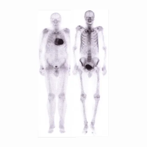
A cardiac scintigraphy with bone-seeking tracers (SPECT) is performed, which demonstrates marked cardiac uptake. This test makes it possible, in many cases, to diagnose ATTR amyloidosis without the need for a biopsy, provided the first-level test results allow it and the scintigraphy result is clearly positive (see Figure).
5. Amyloid typing
This is a fundamental step: knowing whether the amyloid is of AL or ATTR type completely changes the treatment. The most accurate test is mass spectrometry, which precisely identifies the protein responsible for the deposits.
6. Genetic analysis of transthyretin (if the amyloid is ATTR type)
If the SPECT indicates a diagnosis of ATTR amyloidosis, a genetic test is performed to distinguish the hereditary form (ATTRv, linked to a transmissible mutation) from the non-hereditary, age-related form (ATTRwt). This also has implications for the patient's family members.
7. Definitive diagnosis and start of therapy
Once the type of amyloidosis and the organs involved have been established, the multidisciplinary team sets the most appropriate therapy. Starting treatment as soon as possible, before organ damage becomes irreversible, is the factor that most influences long-term outcome.
A single isolated symptom is rarely enough to raise suspicion of amyloidosis. It is the combination of several signs, especially when associated with an unexplained cardiac problem, that should prompt rapid investigation. Every stage of diagnostic delay is a lost opportunity to protect the heart and other organs from damage that, once established, can be difficult to reverse. For this reason, raising awareness among family doctors, specialists, and patients about warning signs remains one of the most powerful tools we have.
In patients with a monoclonal protein, three simple tests
an anticipate the diagnosis of amyloidosis
The role of biomarkers in monitoring patients with MGUS
Anyone living with monoclonal gammopathy of undetermined significance (MGUS) knows that, in most cases, it is a condition to be monitored over time, without the need for immediate treatment. But in a small percentage of patients, the plasma cells underlying the MGUS can produce light chains capable of depositing in the organs in the form of amyloid, giving rise to AL amyloidosis — often years before evident symptoms appear.The good news is that this process leaves measurable traces that can be detected with simple, inexpensive tests already available in routine clinical practice.
Three biomarkers, in particular, make it possible to “listen” early to the three organs most frequently affected:
NT-proBNP → Heart
A hormone released by the heart under stress: it signals possible cardiac involvement even before shortness of breath or fatigue appear.
Proteinuria → Kidneys
The presence of protein in the urine indicates early, often silent, kidney damage.
Alkaline phosphatase → Liver
An increase in this marker can reveal liver involvement before obvious clinical signs emerge.
Why the timing of diagnosis is crucial — Regularly monitoring these three parameters in patients with MGUS — especially when free light chains in the blood are elevated — makes it possible to detect AL amyloidosis while it is still at an early stage, when organ damage is minimal or absent. It is precisely within this time window that early diagnosis makes the greatest difference: starting therapy before the heart, kidneys, or liver have suffered irreversible damage means giving the patient the best chance of responding to treatment and of a long, healthy life.